
Julia Davis / Photos by Matt Hagan
A patch that speaks up when IVs fail
Intravenous therapy is one of the most common medical procedures in the world, with over a billion insertions performed annually. But IV failure, when the needle punctures through a vein and fluid leaks into surrounding tissue, is alarmingly common, especially in newborns. More than 70% of neonates experience IV failure, and because babies can't communicate their discomfort, the problem often goes undetected until serious tissue damage has occurred.
"I've actually talked to mothers of neonates whose children still have scars from the IV failure," says graduate student Surbhi Kakar.

The research team, advised by Professor Dwayne Arola and Dr. Gregory Valentine, a neonatologist at Seattle Children’s, is developing color-changing materials that act as an early warning system for the two most common signs of IV failure: fluid leakage and swelling. One material turns from white to blue the moment it contacts leaked IV fluid. Another is a thin film applied near the IV site that shifts color, from red to green to blue, as the skin beneath it swells. Kakar, who leads the prototyping effort for the swelling design, developed the film.
The goal is to combine both materials into a single patch that integrates into the existing hospital workflow, giving clinical staff a visual signal they can spot across the room. "It's basically to give nurses an extra set of eyes, especially where it's super busy and super under-resourced," says Kakar.
Some hospitals, the team found, didn't even have solid data on how many children were experiencing IV failure because it was so common it went untracked. A low-cost, easy-to-read patch could help change that.
A safer way to see inside
When doctors perform line placement procedures, such as inserting catheters, ports or other instruments into the body, they typically rely on continuous X-ray imaging to see where those instruments are. That means clinicians must wear heavy lead vests, work in expensive specialized suites and accept the cumulative risks of radiation exposure, including increased chances of thyroid cancer and cataracts.
Undergraduates Ryan Parekh and Omar El Souessy wanted to find a better way. Working under Avik Som, an assistant professor in MSE and interventional radiologist at UW Medicine, the two began by simply observing.
"We were told to go in with fresh eyes and see what clinicians were struggling with," says El Souessy. The answer kept coming back to radiation.
Their solution, called MagnaTrace, replaces continuous X-ray imaging with a grid of magnetic sensors. The system tracks the magnetic field of a guidewire as it moves through the body, generating a real-time heat map that shows the instrument's position, no radiation required.

To test whether the sensors could read a magnetic signal through tissue, the team used chicken breast as a stand-in for the human body and demonstrated that the system could detect and locate multiple magnetic sources simultaneously.
The potential impact goes beyond clinician safety. A fully equipped interventional radiology suite can cost upwards of $2 million. Parekh estimates MagnaTrace could bring the cost down to $10,000 to $25,000, making line placement procedures accessible to hospitals with fewer resources.
"It's a lot more portable," says El Souessy. "You can do it bedside if you really want."
The team presented the research at the Society of Interventional Radiologists conference in Toronto in April, and El Souessy plans to continue the research as part of his applied master's at the UW.
Pedaling past carbon fiber
Carbon fiber has long been the gold standard for high-performance cycling shoes. It's stiff, it's light and it transfers power efficiently from foot to pedal. But graduate student Cal Davis thinks the cycling industry has been over-engineering the problem.
Davis is working with Brevay, a Seattle-based cycling footwear company, and adviser Navid Zobeiry to prove that flax fiber can do the job just as well, with a fraction of the environmental impact. Recent biomechanics research suggests that cycling efficiency plateaus after a certain stiffness, and Davis sees that as an opportunity. "As long as we hit a certain stiffness, we should have the same performance as a carbon fiber shoe," they say.
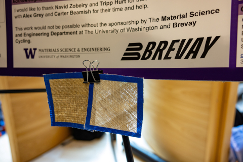
That opens the door for flax. The same plant used to make linen clothing can be processed into fibers that, once set in resin, become surprisingly rigid. Davis is testing flax alongside basalt and hemp fibers, running stiffness and strength tests on prototype outsoles. So far, their prototypes exceed the stiffness threshold needed for performance, though strength requirements still need work before a fully flax outsole is ready for production.
The environmental case is straightforward: flax grows out of the ground and requires far less energy to produce than carbon fiber.
"It's significantly better for the environment," says Davis.
Brevay plans to launch its first road cycling shoe later this year, and Davis's research could help shape what's inside it.
A scaffold for healing
Stem cell therapy holds enormous promise for repairing damaged tissue in the body, from worn-out knee cartilage to fractured bone. But getting the cells to actually do their job is harder than it sounds.
When stem cells are injected into a damaged knee joint, for example, fewer than 5% of those cells typically survive beyond the first few days. Without something to hold them in place, the cells scatter from the injection site, and without the right structural and chemical cues, most die before they can do any good.
Graduate student Jacob Beitzel works in the lab of Miqin Zhang, who holds the Kyocera Corporation Chair in Ceramic Engineering in MSE. Beitzel is developing injectable hydrogels designed to solve that problem.

Made from chitosan, a natural material derived from crustacean shells, the hydrogels are roughly 90% water and act as scaffolds: a supportive structure that holds stem cells at the injury site instead of letting them drift away. The gel components are loaded into a syringe and mixed at the moment of injection, solidifying into a network almost immediately once inside the body. The gels also deliver growth factors that help coax those stem cells into becoming the right type of tissue, whether that's bone, cartilage or something else.
"The key thing is keeping the stem cells where you put them," says Beitzel. His hydrogels have kept cells alive through 14 days in lab testing, with little to no cell death. The gels also showed early signs of bone growth and produced proteins associated with cartilage repair.
What makes the hydrogels especially versatile is that their properties are adjustable. Changing the concentration of the gel changes how stiff it is, so the same basic material can be tailored for soft tissue like cartilage or harder tissue like bone. The gels are also self-healing: if the material is damaged by a sudden twist of the knee, for instance, the bonds reform on their own, eliminating the need for another injection.
Beitzel, who is in his first year of the Ph.D. program after completing his master's at the UW, has been working on the project for nearly three years, starting during his master's.
His next goal is testing in live animal models "to see if it actually can work in a living environment."
If the hydrogels perform as well in living tissue as they have in the lab, they could eventually offer a simpler, less invasive option for people facing joint repair or bone injuries.
Originally published April 30, 2026